How a root canal treatment can save your natural tooth?
Writes: Dr. Elena Saveva
There is no such a thing that looks, feels or functions like your natural teeth. One of the best ways to make sure that you will keep your natural teeth for a long time is to practice good oral hygiene. Brushing your teeth on a regular basis with soft-bristled toothbrush and fluoride toothpaste, flossing between the teeth, using mouthwash after brushing and flossing, eating a healthy diet and limiting sugary food and drinks, along with six-month check-ups at your dentist’s office, can help you keep your teeth for a lifetime.
When a tooth is infected or damaged, it may seem like pulling it is the easiest choice, especially if you are in pain. But missing just a single tooth can have serious consequences, both from a health and quality of life perspective. When possible, you should always consider undergoing root canal treatments to save your teeth. At our clinic, we like to see our patients keeping their natural teeth for as long as possible. When you ask us for advice on what to do with infected tooth, we will give you an outline of why root canal is often the better option compared to any other regarding this issue.
What is root canal treatment and when it is necessary?
In the past, a toothache would have meant extraction and loss of the tooth. Fortunately, as the dental procedures and materials are advancing and are getting better and better every day, we can save teeth with root canal treatment instead. This kind of therapy, also known as endodontic treatment, is dental procedure to treat infection of a tooth’s inner part called tooth pulp.
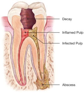
The pulp or the center of the tooth is soft tissue that contains blood vessels, nerves, and connective tissue and is responsible for forming the surrounding dentin and enamel (those are the hard tissues also termed calcified tissues) during tooth development. The pulp receives its nourishment supply from vessels which enter the end of the root. Although the pulp is important during the development of the tooth, it is not necessary for functionality of the tooth. Endodontic treatment is necessary when the pulp becomes inflamed or damaged. The most common reasons for inflammation or infection are deep cavities (caries), deep fillings close to the nerve, repeated dental procedures(for example replacing a large filling), traumatic damage such as cracks or fractures etc.
Root canal treatment in our dental clinic – step by step
1. The first step in the procedure is to take an X-ray to see the shape of the root canals and determine if there are any signs of infection in the surrounding bone. Then we usually use local anesthesia to numb the tooth that is about to be treated and surrounding tissue.
2. Placing the rubber dam
Rubber dam is a thin rubber sheet that is used in a number of dental procedures to keep the tooth dry during treatment and isolate it from the rest of the oral cavity. This allows the root canal treatment to be carried out in a sterile environment, free from contamination by bacteria in saliva or the rest of the mouth. The rubber sheet also protects the oral cavity from foreign objects and chemicals used during the treatment.

3. Creating the access cavity.
The doctor makes an opening (small hole) in the crown of the tooth. The main objective of access cavity preparation is to identify the root canal entrances for subsequent preparation and obturation of the root canal.
4. Identifying all of the tooth’s root canals.
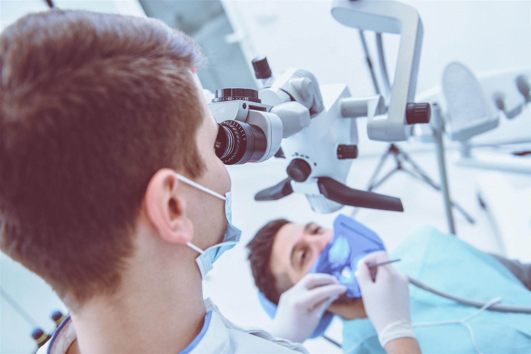
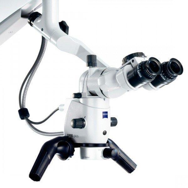
In this stage of the endodontic treatment of considerable help is our dental operating microscope. Canals that are hidden, extremely narrow or unusually positioned become easier to be seen with dental microscope. The superior lightning and the several magnifications available on the dental operating microscope brought many benefits to the therapy.
Some clinical procedures, as endodontic retreatments, removal of separated instruments, intra-canal perforation repairs or working on calcified canals should not even be attempted without the use of the microscope under the risk of heavy tooth structure sacrifice. In our office, we have the most advanced dental microscope available on the market, Zeiss Extraro 300.
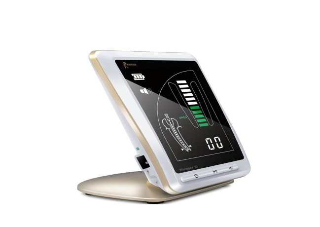
5. Measuring the length of the tooth’s root canal.
The removal of whole pulp tissue, necrotic material and microorganisms from the root canal is essential for endodontic success. This can only be achieved if the length of the root canal is determined. Correct determination of tooth length is a crucial factor for the success of endodontic therapy. X –rays, electronic apex locators and operator's tactile sensation are methods used for determination of root length.
Apex locator is electronic device that uses an electrical circuit through the patient's root canal and oral tissues to determine the location of the apical foramen. It is dominant with precision among the other methods of determining the root canal length.
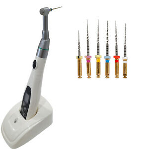
6. Cleaning and shaping the tooth’s root canals.
The main objectives of canal preparation (cleaning and shaping) include:
➢ The removal of vital and necrotic pulp tissue from the root canal space.
➢ The removal of infected hard tissue (dentin)
➢ Removal of microorganisms and their products
➢ Three-dimensional shaping of the canal walls and providing space for placing obturation materials.
To achieve these goals, doctors use endodontic files (manual and rotary) and irrigants (chemical solution with antimicrobial effects). At our dental clinic we use rotary NiTI instruments ,which help us to increase efficacy in this part of root canal treatment and also allow us to treat our patient quickly and comfortable. With a continuous, slow and steady rotation, these instruments that are made of a nickel-titanium alloy are more flexible than the traditional stainless-steel manual files. The flexibility and unique movement of this files allows very thin and curved root canals successfully treated. Activated by a small electric motor, they make the root canal procedure much faster than with conventional files. This does not mean that we have turned our back on manual files, but rather use these new technologies as a supplement to manual files in order to increase the quality of root canal treatment.
7. Root canal obturation.
Root canal obturation is three dimensional filling of the entire root canal system with biologically acceptable materials. Usually, the canal system is obturated with gutta-percha along with a sealing cement.
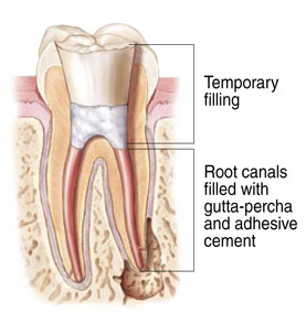
8. Restoration (rebuilding the tooth).
After the root canal obturation is completed and the tooth has “settled down” (no more pain from that tooth), it’s time for post and core buildup. A post and core is a dental restoration used to sufficiently build –up tooth structure for future restoration (crown) when there is not enough structure.The post is a small rod (metal or fiber) that is inserted into the root canal. Before placing the post, we remove a part of the canal filling material creating a space. The post is used to anchor the core and improve the strength of the tooth. A core is built from filling material (composite) around the part of the post that sticks out of the root after the post is cemented into the root. The core is shaped so that it will anchor and retain a crown. The last step of this treatment is making a crown In order to protect treated tooth from possible fracture.
BENEFITS of root canal treatment:
1. Saving your natural teeth
Nothing, not even the most advanced bridges and implants, can truly replace your natural tooth. There are a lot of advantages of saving your tooth with root canal treatment: maintaining your natural smile, efficient chewing, normal biting, and limiting the needs for more costly, ongoing dental work.
2. Elimination of pain and other symptoms
Root canal therapy removes infected tissue to eliminate the source of pain, discomfort and sensitivity to hold and cold.
3. Protection of surrounding tissue and general health
An untreated dental infection can lead to more dangerous conditions, including an abscess or infection that spreads to other areas of the body.
CONSEQUENCES of missing one or more teeth
1. Chewing problems.
When you have a missing tooth, you may not be able to properly chew the food. Food that is not sufficiently chewed can lead to digestive disorders.
2. Bone lost.
Natural teeth are embedded in the jawbone, and stimulate the jawbone through activities such as chewing and biting. When teeth are missing, the alveolar bone, or the portion of the jawbone that anchors the teeth in the mouth, no longer receives the necessary stimulation, and begins to break down, or resorb. This bone loss can lead to jaw shrinking, making it less stable to support the remaining teeth and altering the shape of your face and your smile.
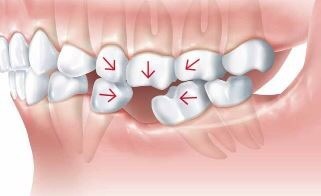
3. Shifting of teeth.
Your teeth naturally support each other, so when one is missing, it can cause adjacent teeth shifting to fill the void left from the missing tooth. This occurrence result in a partial gap and crooked teeth, which are difficult to clean and maintain. This shift in the alignment of your teeth will also have a negative impact on your TMJ.
Schedule your visit today.
Easily access our services via phone or our
online contact form if you have any questions
or would like to schedule an appointment.
Our team will reply to you as soon as possible.